Matteo Fabbri

Research Summary

My research combines the fossil record and developmental biology of modern organisms to understand how evolutionary innovation shaped the origin and radiation of major animal groups alive today.

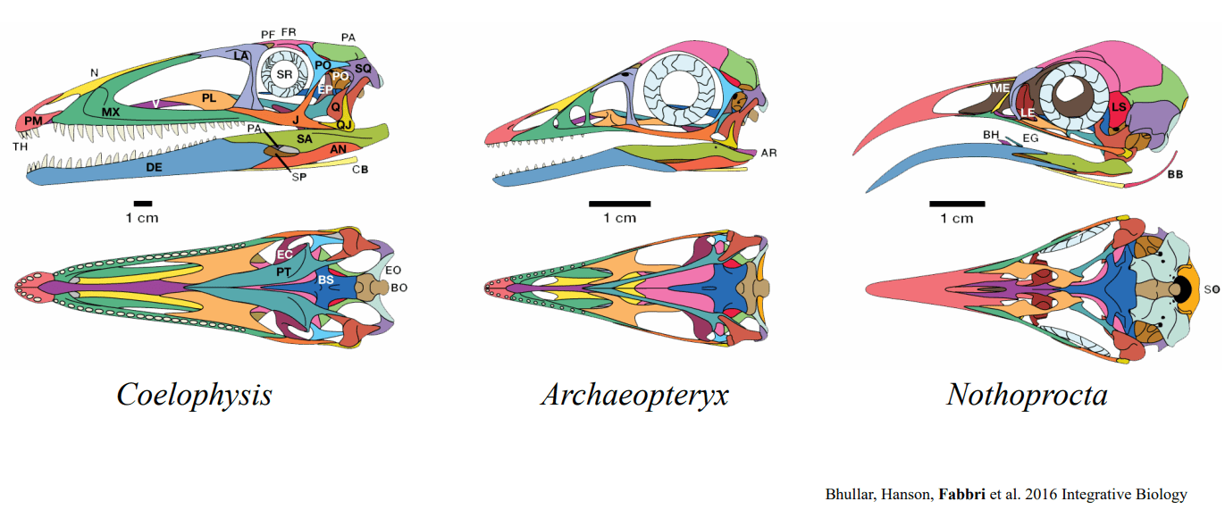
Evolutionary transformation of the skull along the dinosaur-bird transition. Colors indicate the same bone. Image modified from Bhullar et al. (2016)

Why the fossil record?

More than 30.000 species of amniotes (mammals, lizards, turtles, crocodiles, and birds) are alive today. However, their incredible morphological and ecological diversity is the product of hundreds of millions of years of evolution: 99.9% of the species that ever lived on our planet are extinct and can only be studied through the use of fossils.

The fossil record is, therefore, vital to understand the history of amniote groups in deep time, conceptualizing the series of events that favored the divergence of their extant diversity, origin and radiation.

Developmental formation of the head in a mammal embryo (source: gifer.com)

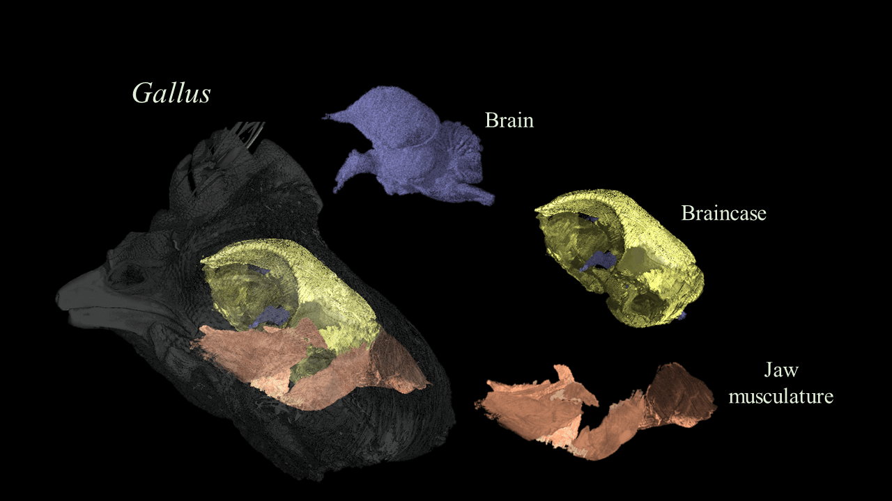
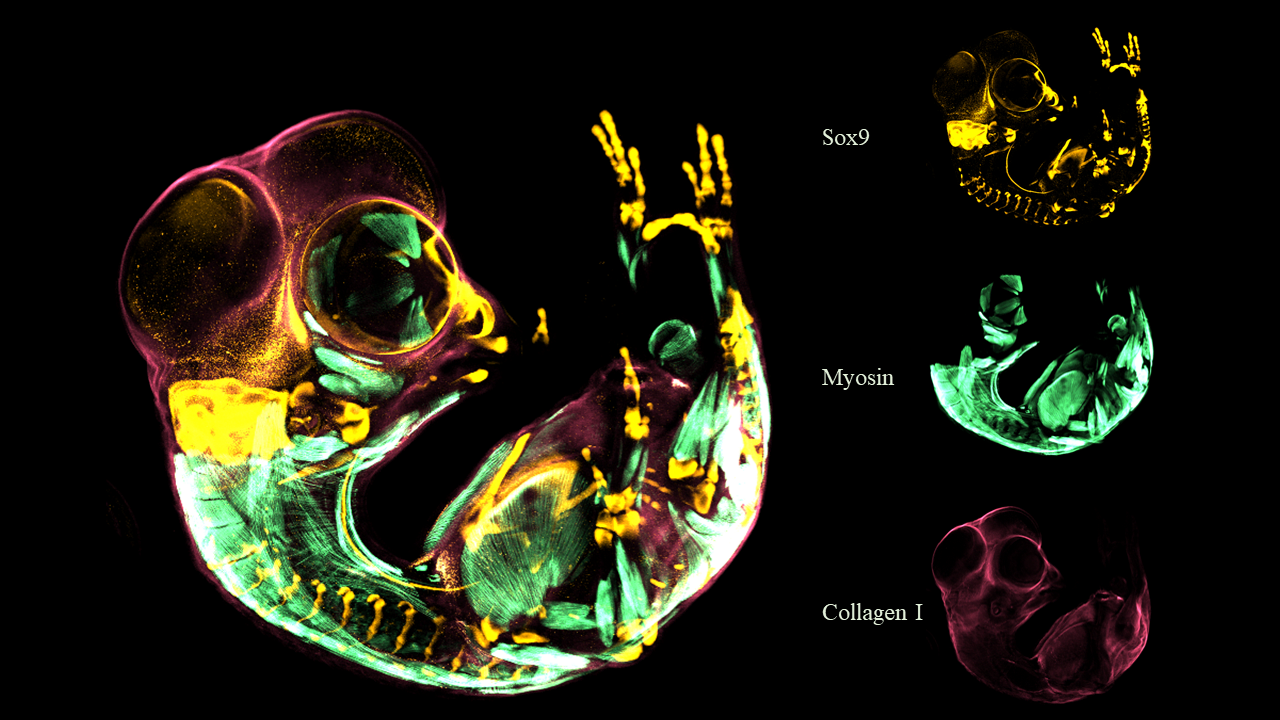
Why the study of embryos?

The geological timespans over which the series of events giving birth and rise to the major amniote clades are recorded is simply too broad and imprecise to approach questions at the mechanistic level necessary to understand the evolutionary process. In order to clarify how certain traits evolved, I study the plasticity of embryos and the developmental pathways characterizing them. At the beginning of development, embryos belonging to different groups are extremely similar to each other in their morphology. It is only later in development that unique traits, such as the bird beak or the mammalian ear, appear.

This opens new avenues for the study of morphology, and in the understanding of constraints and innovations affecting the rise of the major amniote clades.

How can we combine these two sources of data?

Because the fossil record mainly preserves morphological data, I apply cutting-edge imaging techniques, such as micro-CT scan, synchrotron, and CLARITY with immunofluorescence to combine the fossil record with embryological data.

Micro CT scan of a chicken head showing the anatomical relationship between the brain, braincase, and jaw musculature

Micro-CT scanning

X-ray Computed Tomography (CT scanning) allows to obtain anatomical details that are otherwise obscured. These include, but are not limited to, the morphology of the inner skull, such as the palate and the braincase, and the reconstruction of the sensorial capabilities, for example the brain morphology and the inner ear. Additionally, CT scan is an excellent tool for non-invasive virtual dissection of specimens, both extinct and extant.

Virtual dissection of the head of an opossum embryo. The scan was produced with soft tissue staining and synchrotron imaging. The brain is highlighted in blue, the first condensation of the skull in orange, and the mesenchyme in red.

Synchrotron

Because some anatomical details, such as cellular organization and tissue development, are too small to be imaged with standard CT scanning, I use synchrotron imaging. This tool allows to capture anatomical structures at the nano-scale, revealing details that would be impossible to capture with other technologies. My previous data collection was mainly focused on early developmental patterning of embryos and was made possible through the Facilities available at the Argonne National Laboratory.

Zebrafinch embryo imaged whole mount with CLARITY and immunofluorescnce

Clarity and Immunofluorescence

From whales to birds, every single amniote starts development from a single cell, called the zygote. It is only during development that their anatomy appears, leading to the peculiar traits characterizing each amniote groups. While the anatomy of reptiles, birds and mammals is relatively well known from the latest stages of development, the earliest moments of skeletal, muscular, and neurovascular patterning are yet poorly known. Subtle changes happening in the earliest moment of development lead to dramatic changes in the adult morphology of animals. Because of this, I use this staining technique to track the earliest moments of tissue formation and cell aggregation within an evolutionary framework.

Funding

A special thank goes to all Foundations and Institutions that made my research possible over the past years: